Use of Infrared and Ultrasound in Vein Visualization
Infrared and Ultrasound in Vein Visualization refers to the use of the two most popular aides to help clinicians locate veins when placing intravenous (IV) lines or performing blood draws. Infrared devices provide a visual image of the patient’s veins and arteries on their skin, and ultrasounds display these images, and more, on a screen. Both tools can be helpful, but despite the undisputed benefits of vein visualization technology, it should always be utilized in conjunction with a thorough patient examination prior to beginning any procedure, including visually assessing the patient’s physical condition and medical history. Read more to learn about the use of infrared devices and ultrasound equipment in vein visualization and their importance in safe and effective vascular access.
What is Vein Visualization?
Vein visualization describes the method of using advanced technology to display or project images and videos in real time to help identify the location of veins. In addition to the location of veins, the more advanced vein visualization tools can also confirm the exact location of the needle to verify it is in the right place (i.e., vein vs artery, depending on the type of vascular access device being placed). With the right training, needle visualization is extremely valuable in preventing IV medications from being infused incorrectly, which can be a cause of both minor and major injuries to the patient.
Why is Vein Visualization Important?
Vein visualization is a very helpful and important tool that should be used in the placement of intravenous (IV) lines because it enables clinicians to see exactly where a vein is to access it successfully on the first attempt. This reduces the number of attempts to start an IV, which decreases risk of infections, complications, and patient pain and discomfort.
For certain patients, such as those with kidney disease and renal failure, vein preservation (limiting vascular access and attempts to save veins for future use) is extremely important. Failed attempts and medications infused in the wrong space may permanently damage a vein, and as such, starting IVs on the first attempt is important not just for patient comfort, but to ensure safe and viable intravenous sites for current and future therapy.
What Factors Make Vein Visualization Difficult?
There are many factors that may make both vein visualization and the process of starting an IV more difficult without the assistance of a vein finder tool. Some examples include:
Darker Skin Tones – Veins can be harder to see on darker or tattooed skin, so clinicians must rely more heavily on palpation and other methods to visualize veins.
Higher BMI – When a patient has excess weight, veins sit deeper under the skin, making them harder to palpate, visualize, and access.
Medical History – Some patients have medical conditions or have had procedures that can cause a DIVA (difficult IV access) condition due to vein deterioration, stenosis, or complete occlusion of the vessel. Examples include chemotherapy, diabetes mellitus, lack of weight bearing exercise, and vascular conditions.
Dehydration – When patients are dehydrated, they have less total blood volume, which makes veins smaller and harder to access.
Hypotension (Low Blood Pressure) – There are many causes of low blood pressure, including heart conditions, medications, dehydration, and more. Low blood pressure can cause veins to flatten, making them more difficult to access.
Vein Characteristics – Some people naturally have smaller veins, poor circulation, or other factors that make it more difficult to find a vein.
Anxiety – Anxiety activates the sympathetic nervous system, which causes vasoconstriction (narrowing or constricting of blood vessels/veins) and makes them harder to locate and access.
What types of Vein Finder Devices can be used to Visualize Veins?
There are several types of Vein Finder Devices that clinicians can use to help locate and visualize veins. Some devices use infrared technology while others use high-frequency sound waves to display images of veins. As is often the case, the vein visualization devices or aides that offer the greatest detail and accuracy are more expensive and require extensive training, precepting and practice to gain competency.
Use of Infrared and Ultrasound in Vein Visualization
How are Infrared Vein Finders used to Visualize Veins?
One of the most common types of vein finders used to visualize veins are infrared devices. These devices use hemoglobin molecules in the blood vessels to identify and project an image onto the skin that shows where the veins are located. Infrared vein finders are relatively inexpensive, although they can vary in price depending on their features. Infrared vein finders are generally easy to learn how to use and can be helpful in mapping location and size of veins in a patient.

Infrared Device and vein visualization

Infrared Device to visualize veins
While these devices can be helpful in many cases such as blood draws and placing peripheral IVs (PIVs), they have some limitations. For example, infrared visualization devices do not work well in obese patients (veins are deeper and harder to detect), tattooed patients, or those with African American or Asian ethnicity (projected images are more diffcult to visualize on darker pigmented skin). Additionally, the image of the vessel that’s portrayed on the skin can easily be distorted if the distance away from the surface of the skin is not held correctly. Finally, infrared devices only penetrate the skin a few millimeters, so deeper vessels cannot be visualized.
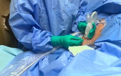
How are Ultrasounds used to Visualize Veins?
Ultrasound imaging is another type of technology that can be used to visualize veins. These devices use sound waves to identify blood vessels as well as other structures and offer many advantages over vein finders that use infrared technology. Because ultrasounds use sound waves to display what is occurring under the skin, the accuracy and detail is not impacted by darker skin tones, tattoos or other scenarios that may impede the ability to visualize veins. Additionally, because ultrasounds can locate deeper veins than infrared technology, they are particularly helpful in both blood draws and placing vascular access devices in veins that are deeper in the tissue and not able to be visualized by with infrared technology.

Ultrasound Device
to visualize veins

Ultrasound Device
showing vein visualization
An important advantage of ultrasound imaging over infrared technology is the ultrasound’s ability to provide “Needle Visualization,” which shows exactly what is taking place beneath the skin. Needle visualization enables clinicians to not only see where the vein is, but also exactly where the needle is. This ensures that the needle enters the correct anatomy (i.e., vessel vs artery) and allows for single punctures of vessels rather than a “double wall” puncture where the needle travels through both walls of the vessel rather than staying inside it. Double wall punctures contribute to bruising as well as possible leaking of the administered medication outside the vessel. Leaking of certain medications such as vesicants (e.g., harsh medications, which include vasopressors, some antibiotics such as vancomycin, and chemotherapy agents, to name a few) outside the vein can be dangerous to the patient, and can even cause permanent damage in some scenarios. With proper training, the percentage of single stick success is very high, approaching 100%. For patients who have needle fear from previous multiple painful attempts, this is an important factor in patient satisfaction.
Read more about IV Infiltration and Extravasation prevention and management HERE
While ultrasounds are the best tools to assist in vein visualization, they are more expensive to purchase, repair, and maintain, they require a higher level of training for proficiency, and even more for expertise. As a result, even though ultrasound guidance is a significantly more reliable method, many facilities do not have the resources to buy the equipment or provide this extensive training. One of the advantages of being skillfully specialized in vascular access is it ensures the higher level of expert training which leads to greater success.
Use of Infrared and Ultrasound in Vein Visualization
Vein visualization is an important part of IV access. While there are many tools to aid in vein visualization, ultrasound guidance is the most accurate and reliable method. Ultrasounds can be used where traditional landmark or infrared guidance falls short, making them a more consistent and reliable alternative to other methods. These benefits ensure accurate and quality vascular access on the first try to reduce the number of sticks and the risk of complications and infections, and further, to improve patient satisfaction and promote vein preservation.
Learn More about Needle Visualization
Our Director of Research and Development talks about our extensive training and needle visualization in this video, click the play button to hear what he has to say.
Vascular Access Education and Training
Comprehensive vascular access education and training that includes didactic classroom education, hands-on simulation and precepting are important components of becoming not just proficient, but competent, in placing ultrasound-guided vascular access devices. Vascular Wellness offers Education and Training courses with the opportunity to earn CEs and achieve clinical competency on ultrasound-guided PIVs, Midlines, PICCs, and Small Bore (Internal Jugular and Mid-Thigh Femoral) Central Venous Catheter (CVC or Central Line) insertion. We also offer courses on the identification, care and maintenance of vascular access lines, and more. All courses are taught by Vascular Access Board Certified (VA-BC), practicing nurse clinicians who regularly place lines and precept and train our nurses.
Interested in Learning more about Vascular Wellness Education and Training Courses?
Click HERE to read more about Education and Training.
Curious about Vein Preservation for Kidney Disease and Renal Failure Patients?
Learn more about the vital role vein preservation plays in patients with kidney disease and renal failure. Click HERE to read about Vein Preservation and Kidney Disease.
Vascular Access Patient Cases:
Clinical Cases from the Front Lines of Vascular Access Care

We place the right line at the right time, the first time.
Call us at 877-284-4435 or email us to learn more.
No upfront cost or commitment to get started.
If you require Vascular Access or want to learn more, speak to the team at Vascular Wellness today.
For the latest articles and insights, follow us on LinkedIn, Facebook, Twitter, YouTube, and Instagram.
Vascular Wellness provides:
(1) Comprehensive vascular access services to Mississippi, North Carolina, Ohio, Oklahoma, Pennsylvania, South Carolina, and Virginia; and
(2) Customized vascular access services to Arkansas, Delaware, Georgia, Tennessee, and West Virginia; and
(3) Support vascular access services to Kentucky.